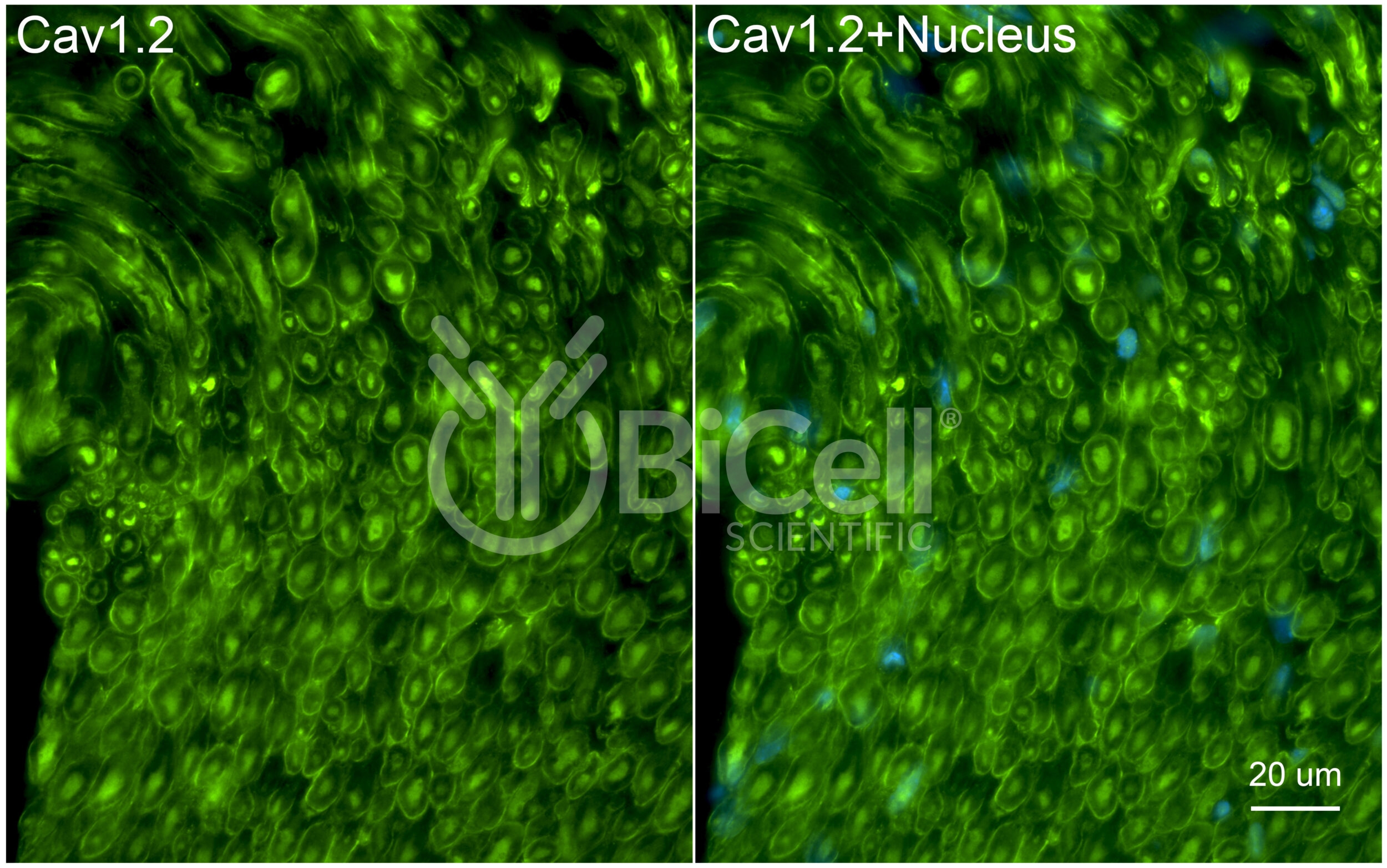
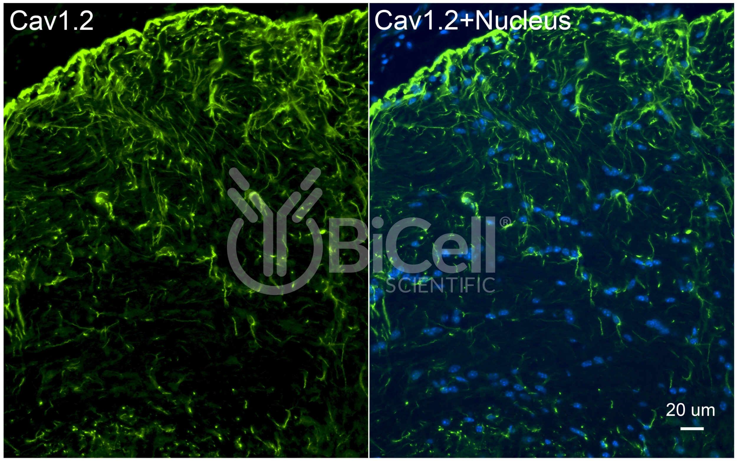
CACNA1C (Cav1.2) Antibody
Description
Anti-CACNA1C (Cav1.2) antibody is validated on mouse tissue and recommended for immunofluorescence labeling, IHC, or western blot of materials from rodent and human tissues. Calcium channel, voltage-dependent, L type, alpha 1C subunit (also known as Cav1.2) is a protein that is encoded by the CACNA1C gene in human. The alpha-1 subunit consists of 24 transmembrane segments and forms the pore through which ions pass into the cell. The calcium channel consists of a complex of alpha-1, alpha-2/delta, beta, and gamma subunits in a 1:1:1:1 ratio. Cav1.2 is widely expressed in the smooth muscle, pancreatic cells, fibroblasts, and neurons. In the heart, Cav1.2 mediates L-type currents, which causes calcium-induced calcium release from the ER Stores via ryanodine receptors.
| Application: | Immunofluorescence, Immunohistochemistry, Western Blot |
|---|---|
| Clonality: | Polyclonal |
| Concentration: | 0.25 mg/ml |
| Conjugation: | Unconjugated |
| Host: | Rabbit |
| Immunogen: | Synthetic peptide (17-aa) derived from the N-terminal region of mouse Cav1.2 protein |
| Isotype: | IgG |
| Purification: | Affinity Chromatography |
| Reactivity: | Human, Mouse, Rat |
| Species Homology: | Synthetic peptide sequence is identical to rat sequence (showing 94.1% homology to human sequence) |
| Storage: | -20°C |
| Storage Buffer: | PBS, pH 7.2, 0.1% Sodium Azide |


