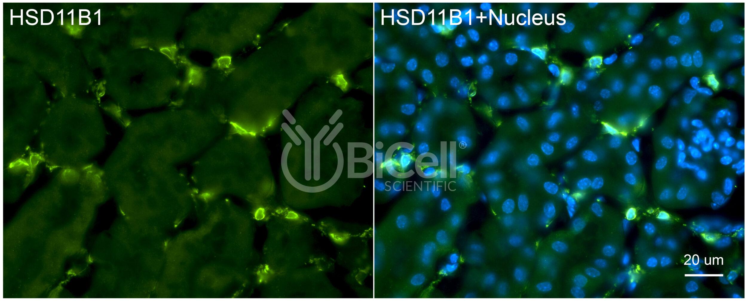
Anti-11-beta-HSD1 (HSD11B1) antibody is validated on mouse tissue and recommended for immunofluorescence labeling, IHC, or western blot of materials from human and rodent tissues.
11-beta-Hydroxysteroid dehydrogenase type 1 (HSD1), also known as cortisone reductase, is an NADPH-dependent enzyme that is encoded by the HSD11B1 gene in human. 11-beta-HSD1 reduces cortisone to the active hormone cortisol that activates glucocorticoid receptors. 11-beta-HSD1 is highly expressed in key metabolic tissues including liver, adipose tissue, and the central nervous system.
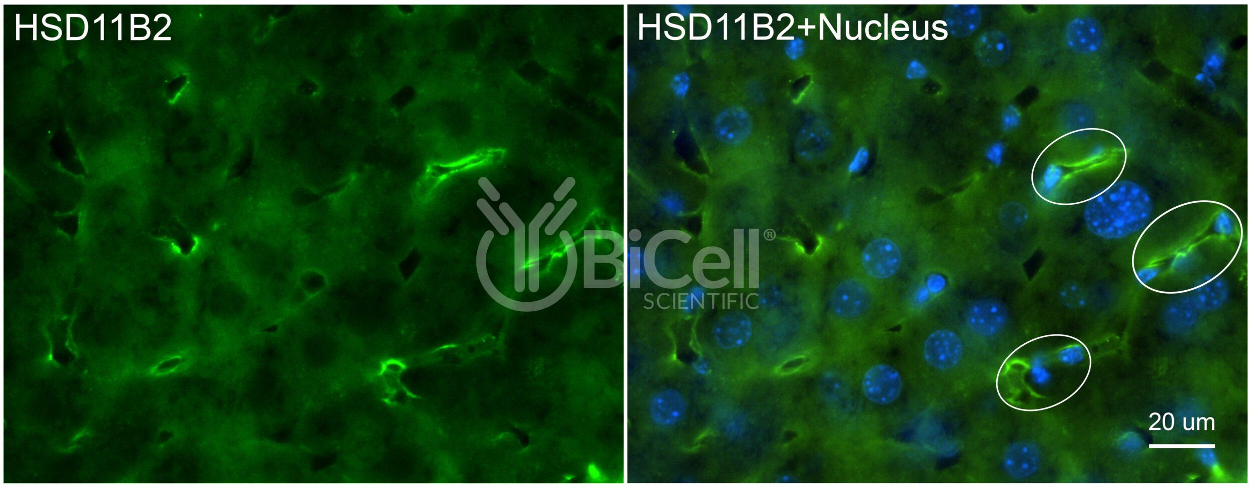
Anti-11-beta-HSD2 (HSD11B2) antibody is validated on mouse tissue and recommended for immunofluorescence labeling, IHC, or western blot of materials from human and rodent tissues.
Corticosteroid 11-?-dehydrogenase isozyme 2, also known as 11-?-hydroxysteroid dehydrogenase 2, is an enzyme that is encoded by the HSD11B2 gene in human. HSD11B2 is an NAD+-dependent enzyme expressed in aldosterone-selective epithelial tissues such as the kidney, colon, salivary and sweat glands. In these tissues, HSD11B2 oxidizes the glucocorticoid cortisol to the inactive metabolite cortisone, thus preventing illicit activation of the mineralocorticoid receptor.
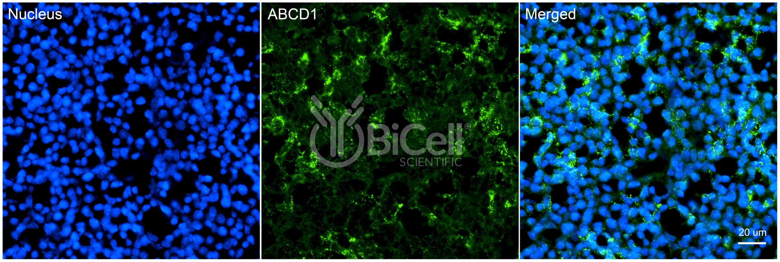
Anti-ABCD1 (human) antibody is recommended for immunofluorescence labeling, IHC, or western blot of materials from human tissues.
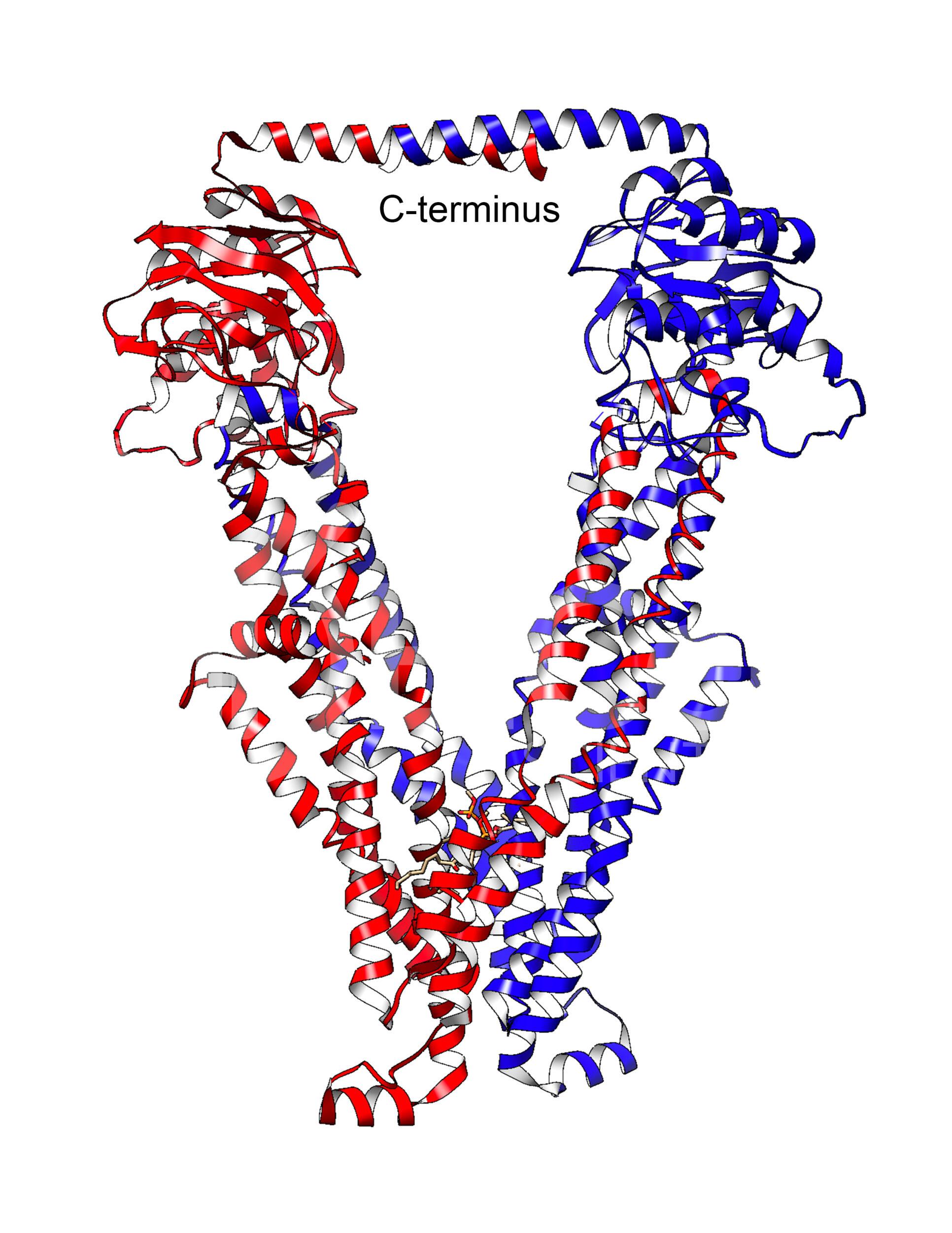
ATP binding cassette subfamily D member 1 is a member of the superfamily of ATP-binding cassette (ABC) transporters, which is encoded by the ABCD1 gene in human. ABCD1 is involved in peroxisomal import of fatty acids and/or fatty acyl-CoAs in the organelle. Defects in this gene have been identified as the underlying cause of adrenoleukodystrophy, an X-chromosome recessively inherited demyelinating disorder of the nervous system.
The epitope sequence used to make this product is homologous to the epitope sequence used in product #21101.
Anti-ABCD1 antibody is validated on mouse tissue and recommended for immunofluorescence labeling, IHC, or western blot of materials from rodent tissues.
ATP binding cassette subfamily D member 1 is a member of the superfamily of ATP-binding cassette (ABC) transporters, which is encoded by the ABCD1 gene in human. ABCD1 is involved in peroxisomal import of fatty acids and/or fatty acyl-CoAs in the organelle. Defects in this gene have been identified as the underlying cause of adrenoleukodystrophy, an X-chromosome recessively inherited demyelinating disorder of the nervous system.
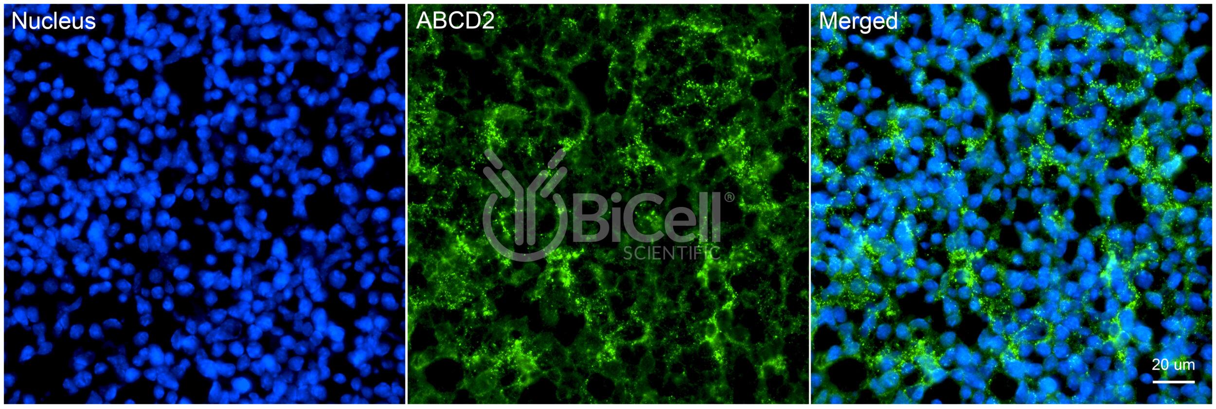
Anti-ABCD2 antibody is validated on mouse tissue and recommended for immunofluorescence labeling, IHC, or western blot of materials from rodent tissues.
ATP binding cassette subfamily D member 2 is a member of the superfamily of ATP-binding cassette (ABC) transporters, which is encoded by the ABCD2 gene in human. ABCD2 is involved in peroxisomal import of fatty acids and/or fatty acyl-CoAs in the organelle. Mutations in this gene have been found in patients with adrenoleukodystrophy, a severe demyelinating disease.
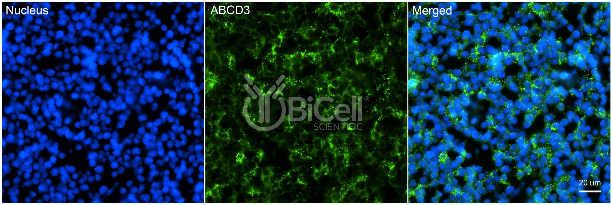
Anti-ABCD3 antibody is validated on mouse tissue and recommended for immunofluorescence labeling, IHC, or western blot of materials from human and rodent tissues.
ATP binding cassette subfamily D member 3 is a member of the superfamily of ATP-binding cassette (ABC) transporters, which is encoded by the ABCD3 gene in human. ABCD3 is involved in peroxisomal import of fatty acids and/or fatty acyl-CoAs in the organelle. Mutations have been associated with some forms of Zellweger syndrome, a heterogeneous group of peroxisome assembly disorders.
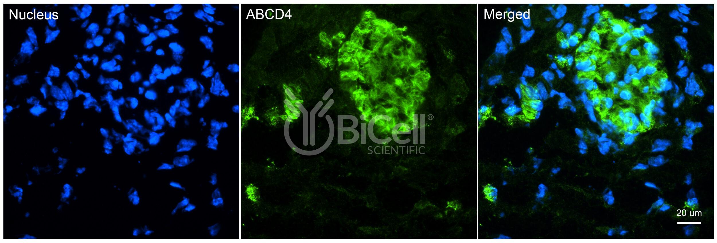
Anti-ABCD4 antibody is validated on mouse tissue and recommended for immunofluorescence labeling, IHC, or western blot of materials from rodent tissues.
ATP binding cassette subfamily D member 4 is a member of the superfamily of ATP-binding cassette (ABC) transporters, which is encoded by the ABCD4 gene in human. ABCD4 is involved in peroxisomal import of fatty acids and/or fatty acyl-CoAs in the organelle. ABCD4 may function as a heterodimer for other peroxisomal ABC transporters and, therefore, may modify the adrenoleukodystrophy phenotype.
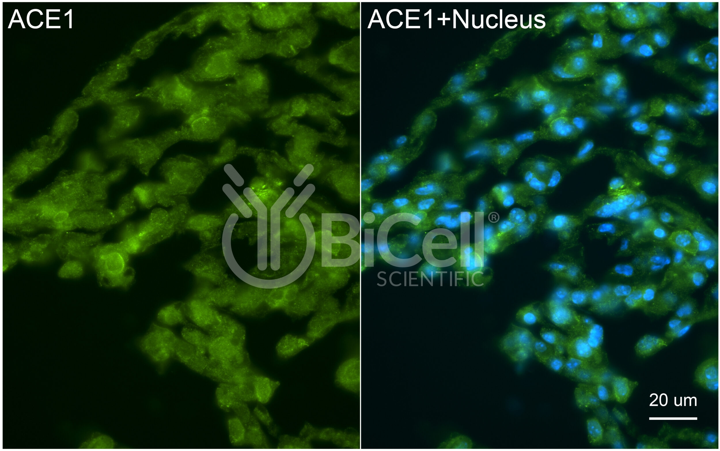
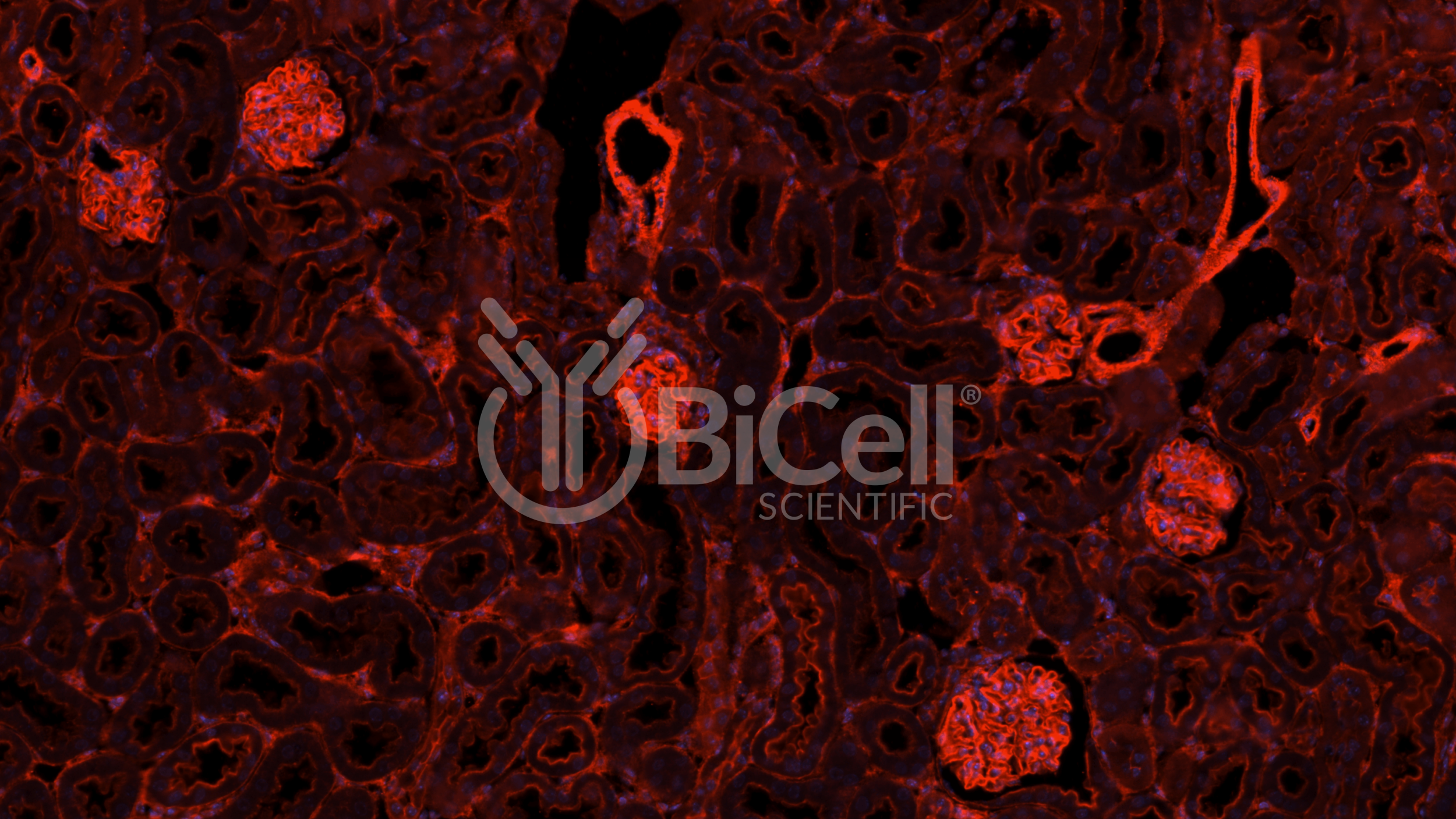
Anti-ACE1 (ACE or CD143) antibody is validated on mouse tissue and recommended for immunofluorescence labeling, IHC, or western blot of materials from human and rodent tissues.
Angiotensin-converting enzyme (ACE, ACE1 or CD143) is encoded by the ACE gene in human. ACE is a central component of the reninangiotensin system (RAS), which controls blood pressure by regulating the volume of fluids in the body. It converts the hormone angiotensin I to the active vasoconstrictor angiotensin II.
ACE is a single-pass type I membrane protein. The extracellular domain of ACE can be cleaved from the transmembrane domain to circulate as soluble ACE or sACE. The ACE gene encodes two isozymes. The somatic isozyme, expressed in the endothelium of many epithelial tissues, has longer extracellular domain, whereas the germinal isozyme, expressed only in sperm, has truncated extracellular domain.
This antibody was raised against the extracellular domain of somatic ACE isozyme and can recognize both soluble and membrane bound ACE.
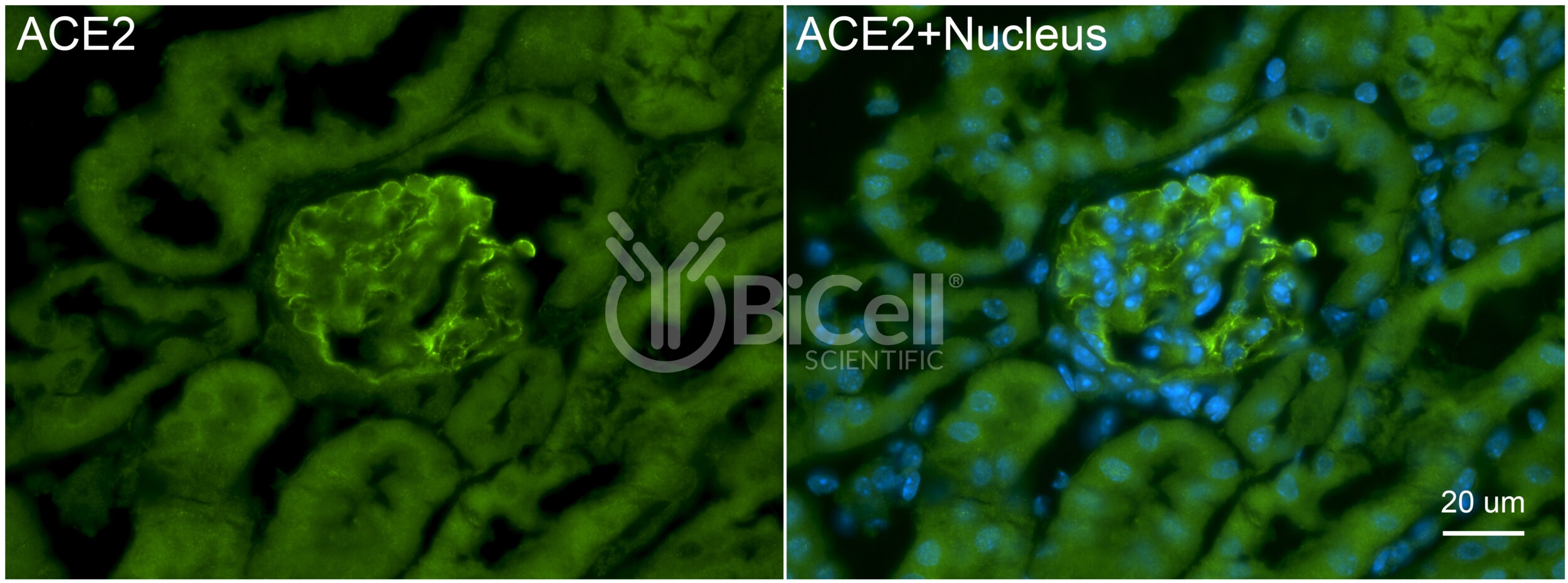
Anti-ACE2 antibody is validated on mouse tissue and recommended for immunofluorescence labeling, IHC, or western blot of materials from human and rodent tissues.
Angiotensin-converting enzyme 2 (ACE2) is an enzyme that is encoded by the ACE2 gene in human. ACE2 is a single-pass type I membrane protein. The extracellular domain of ACE2 can be cleaved from the transmembrane domain by ADAM17. The resulting cleaved protein is known as soluble ACE2 or sACE2. sACE2 lowers blood pressure by catalyzing the hydrolysis of angiotensin II (a vasoconstrictor peptide) into angiotensin (17) (a vasodilator). sACE2 counters the activity of the related angiotensin-converting enzyme 1 (ACE1) by reducing the amount of angiotensin-II and increasing angiotensin (1-7).
ACE2 also serves as the receptor for some coronaviruses, including HCoV-NL63, SARS-CoV, and SARS-CoV-2 (Covid-19). The SARS-CoV-2 spike protein itself is known to damage the epithelium via downregulation of ACE2.
This antibody was raised against the extracellular domain of ACE2 and can recognize both soluble and membrane bound ACE2.
Anti-Actinin alpha-4 (ACTN4) antibody is validated on mouse tissue and recommended for immunofluorescence labeling, IHC, or western blot of materials from rodent and human tissues.
Actinin alpha-4 is encoded by the ACTN4 gene in human. Actinin alpha-4 is an actin-binding protein. In nonmuscle cells, actinin alpha-4 is concentrated along the microfilament bundles near the adherens junctions.
Actinin alpha-4 contains actin binding domain, spectrin domain and EF-hand domain.
Actinin alpha-4 is highly expressed by the glomerular podocytes. Mutations in actinin alpha-4 cause focal segmental glomerulosclerosis (FSGS) (OMIM 603278).
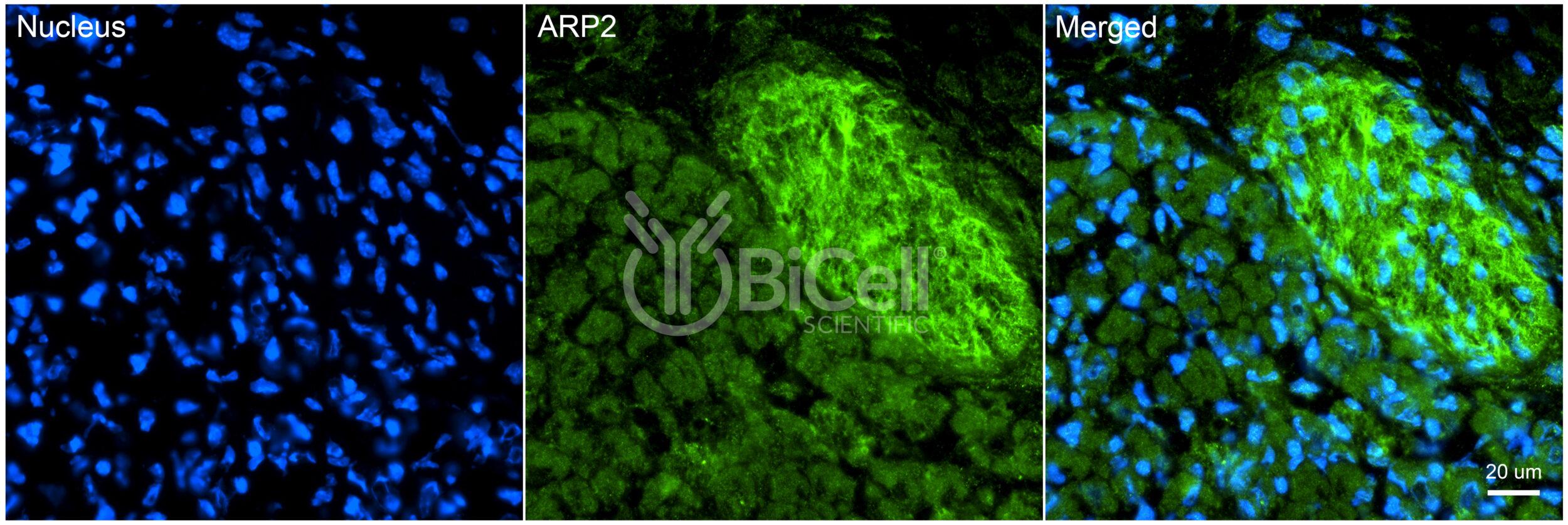
Anti-ACTR2 (ARP2) antibody is validated on mouse tissue and recommended for immunofluorescence labeling, IHC, or western blot of materials from rodent and human tissues.
Actin-related protein 2 is an actin binding protein that is encoded by the ACTR2 gene in human. ARP2 is a component of the ARP2/3 complex. This complex is located at the cell surface and is essential to cell shape and motility through lamellipodial actin assembly and protrusion.
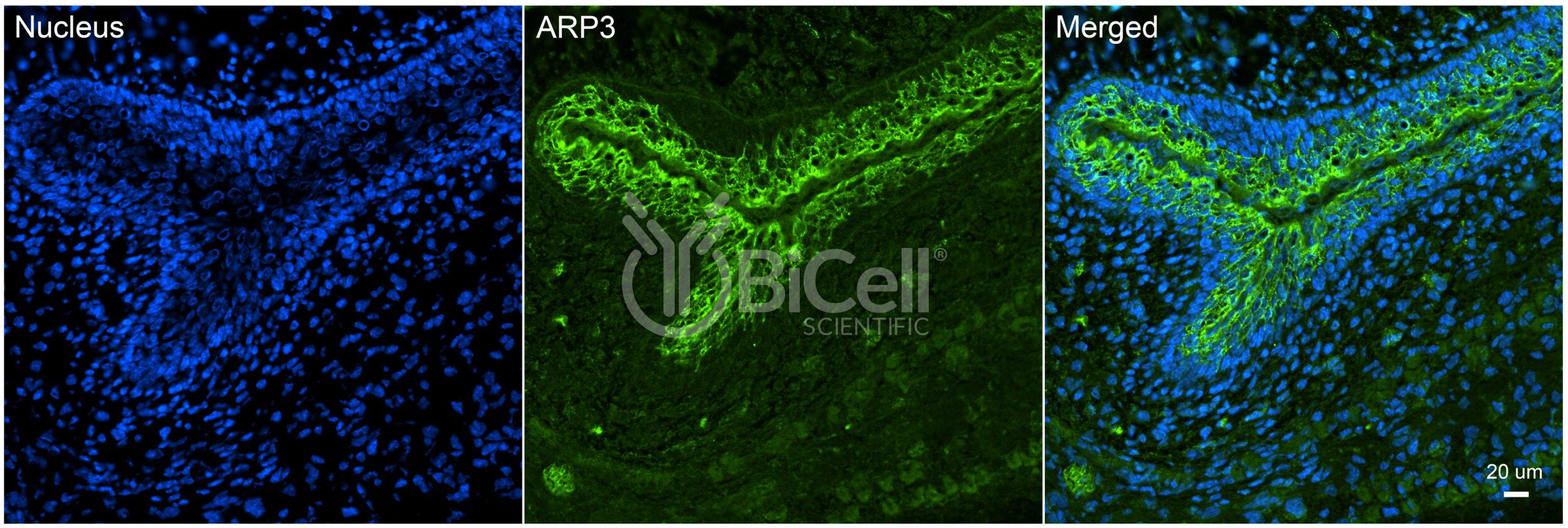
Anti-ACTR3 (ARP3) antibody is validated on mouse tissue and recommended for immunofluorescence labeling, IHC, or western blot of materials from rodent and human tissues.
Actin-related protein 3 is an actin binding protein that is encoded by the ACTR3 gene in human. ARP3 is a component of the ARP2/3 complex. This complex is located at the cell surface and is essential to cell shape and motility through lamellipodial actin assembly and protrusion.
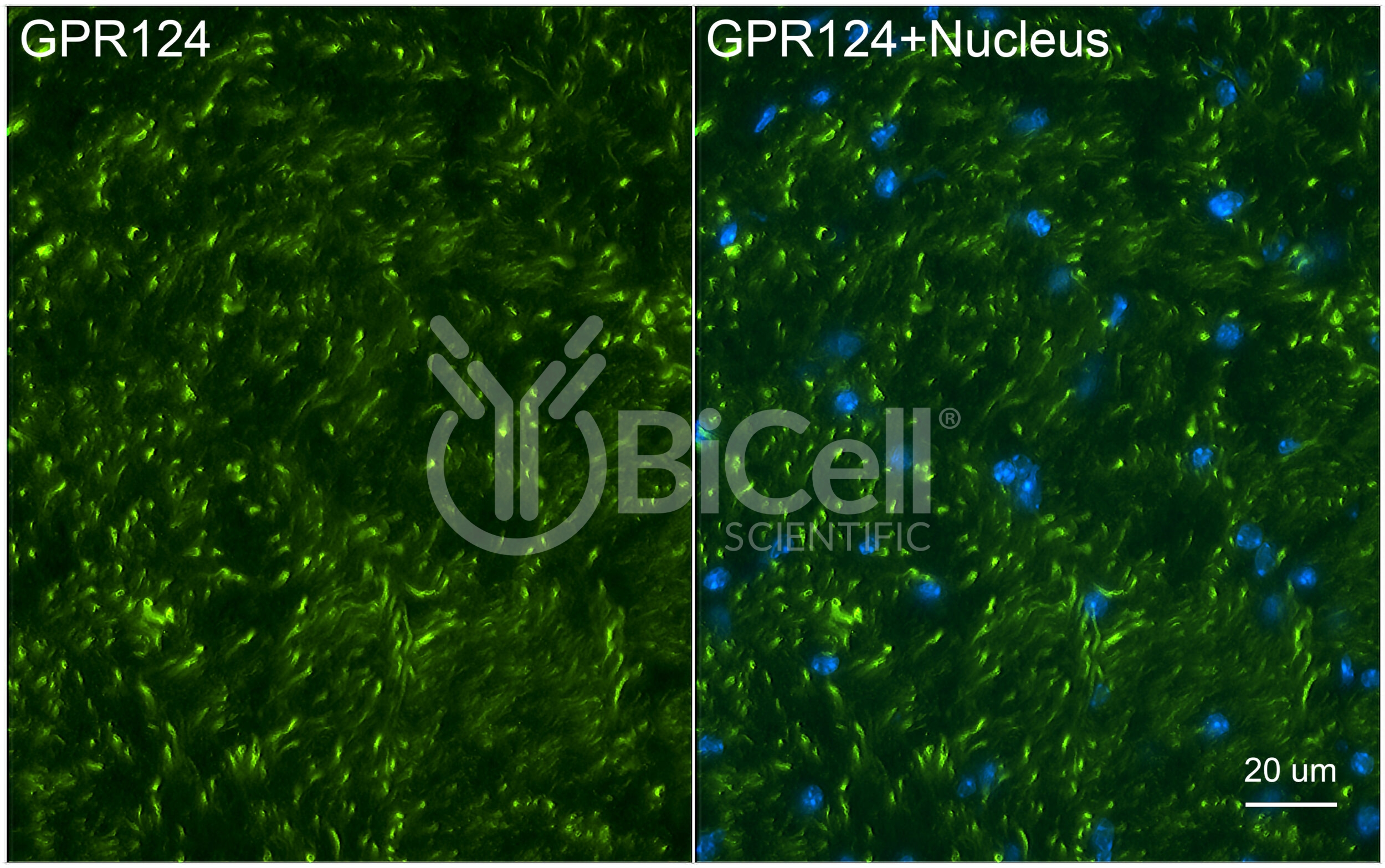
Anti-ADGRA2 (GPR124) antibody is validated on mouse tissue and recommended for immunofluorescence labeling, IHC, or western blot of materials from rodent and human tissues.
Adhesion G protein-coupled receptor A2 (ADGRA2), also known as G-protein coupled receptor 124 (GPR124), is encoded by the ADGRA2 gene in human. ADGRA2 is a member of the adhesion-GPCR family of receptors. Family members are characterized by an extended extracellular region with a variable number of protein domains coupled to a TM7 domain via a domain known as the GPCR-Autoproteolysis INducing (GAIN) domain. ADGRA2 has been shown to interact with DLG1 and is involved in the Wnt/?-catenin signaling pathway along with RECK.
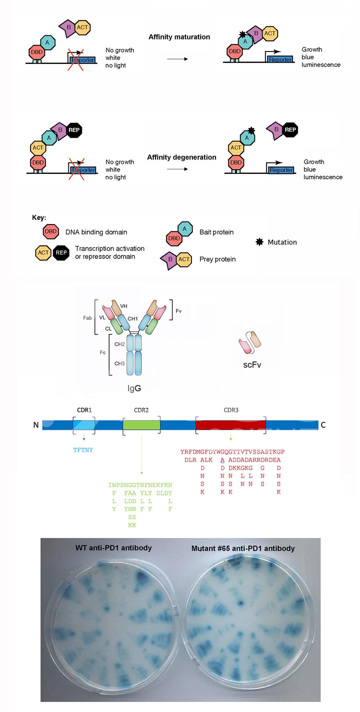
Enhance your research with our Custom Antibody Services, offering tailored Affinity Maturation and Degeneration solutions for precise, high-affinity antibodies. Elevate your project today.

